Dental-system
 Jatin Yadav
Create a AI-Powered Dental Clinic Operating System as centralized SaaS platform


Comments (0)
Sign in to leave a comment
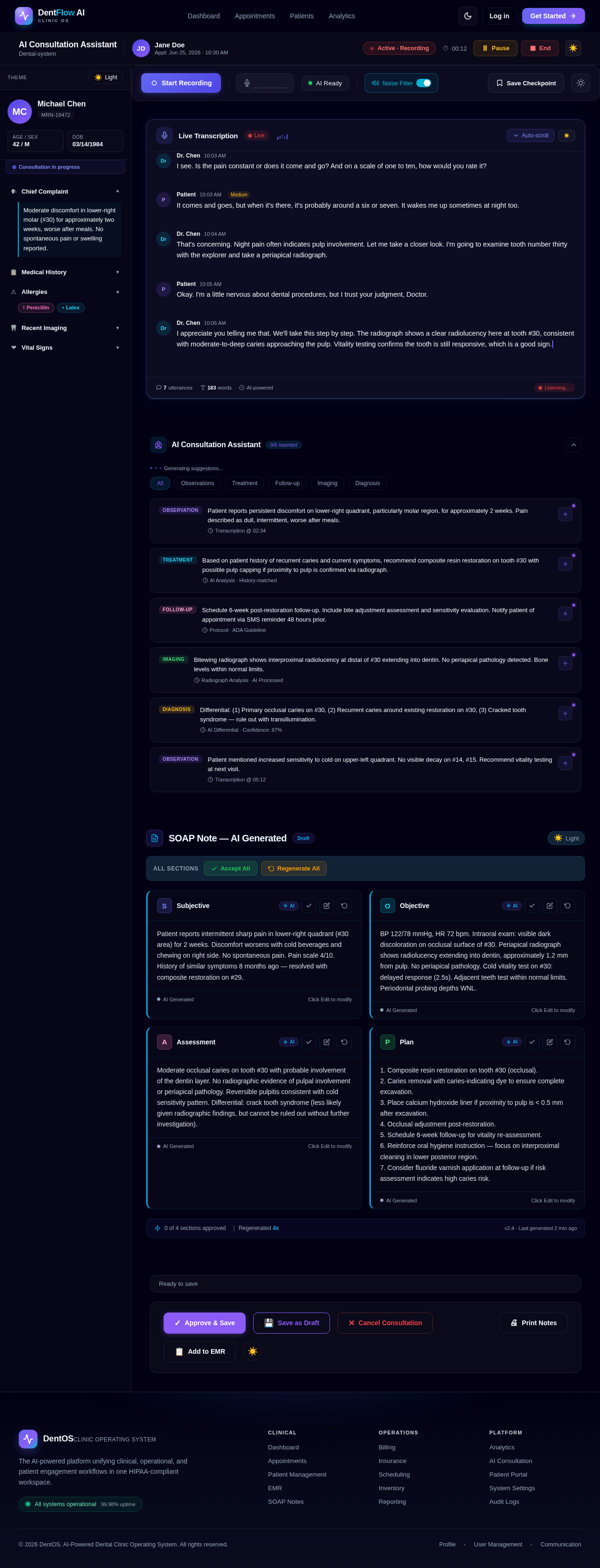
AI Consultation Assistant
Dental-systemPatient reports persistent discomfort on lower-right quadrant, particularly molar region, for approximately 2 weeks. Pain described as dull, intermittent, worse after meals.
Transcription @ 02:34Based on patient history of recurrent caries and current symptoms, recommend composite resin restoration on tooth #30 with possible pulp capping if proximity to pulp is confirmed via radiograph.
AI Analysis · History-matchedSchedule 6-week post-restoration follow-up. Include bite adjustment assessment and sensitivity evaluation. Notify patient of appointment via SMS reminder 48 hours prior.
Protocol · ADA GuidelineBitewing radiograph shows interproximal radiolucency at distal of #30 extending into dentin. No periapical pathology detected. Bone levels within normal limits.
Radiograph Analysis · AI ProcessedDifferential: (1) Primary occlusal caries on #30, (2) Recurrent caries around existing restoration on #30, (3) Cracked tooth syndrome — rule out with transillumination.
AI Differential · Confidence: 87%Patient mentioned increased sensitivity to cold on upper-left quadrant. No visible decay on #14, #15. Recommend vitality testing at next visit.
Transcription @ 05:12SOAP Note — AI Generated
DraftPatient reports intermittent sharp pain in lower-right quadrant (#30 area) for 2 weeks. Discomfort worsens with cold beverages and chewing on right side. No spontaneous pain. Pain scale 4/10. History of similar symptoms 8 months ago — resolved with composite restoration on #29.
BP 122/78 mmHg, HR 72 bpm. Intraoral exam: visible dark discoloration on occlusal surface of #30. Periapical radiograph shows radiolucency extending into dentin, approximately 1.2 mm from pulp. No periapical pathology. Cold vitality test on #30: delayed response (2.5s). Adjacent teeth test within normal limits. Periodontal probing depths WNL.
Moderate occlusal caries on tooth #30 with probable involvement of the dentin layer. No radiographic evidence of pulpal involvement or periapical pathology. Reversible pulpitis consistent with cold sensitivity pattern. Differential: crack tooth syndrome (less likely given radiographic findings, but cannot be ruled out without further investigation).
1. Composite resin restoration on tooth #30 (occlusal). 2. Caries removal with caries-indicating dye to ensure complete excavation. 3. Place calcium hydroxide liner if proximity to pulp is < 0.5 mm after excavation. 4. Occlusal adjustment post-restoration. 5. Schedule 6-week follow-up for vitality re-assessment. 6. Reinforce oral hygiene instruction — focus on interproximal cleaning in lower posterior region. 7. Consider fluoride varnish application at follow-up if risk assessment indicates high caries risk.
No comments yet. Be the first!